

Suppressed acetyl-CoA production promotes mitochondrial health during liver regeneration.
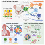
During liver injury and regeneration, fatty acids from adipose tissues transit to the liver to fuel mitochondrial beta oxidation in WT hepatocytes, which outcompete ETC-mutant hepatocytes. In the absence of a functional ETC, fatty acid buildup reduces acetyl-CoA through induction of PDK4. PDK4 inhibition restores flexibility for acetyl-CoA generation, allowing ETC-mutant hepatocytes to proliferate. [Figure created with BioRender.com]
In the realm of liver regeneration, a new study titled “Metabolic Inflexibility Promotes Mitochondrial Health During Liver Regeneration” is shedding light on the intricate processes that maintain tissue health and promote recovery after damage. This research unveils a fascinating mechanism involving mitochondrial function and metabolic pathways that ensure the proliferation of healthy liver cells while limiting the growth of damaged ones.
Key Insights from the Study
Selective Mitochondrial Health: During liver damage, ensuring the proliferation of healthy tissue without spreading diseased cells is crucial. The study by Wang et al. identifies how selective pressure on mitochondria helps achieve this. In conditions like cirrhosis, mutations in the mitochondrial genome can impair the electron transport chain (ETC), which is vital for cellular respiration. Although this dysfunction isn’t directly lethal, it gives affected cells a competitive disadvantage, reducing their metabolic flexibility and ability to proliferate.
The Role of ETC Dysfunction: The research highlights that during liver regeneration, hepatocytes (liver cells) with a dysfunctional ETC struggle to proliferate. This inability to generate acetyl-CoA from peripheral fatty acids through mitochondrial β-oxidation forces a dependence on ETC-functional mitochondria. The accumulation of fatty acids and the suppression of alternative pathways for acetyl-CoA production (from pyruvate or acetate) further impede the proliferation of ETC-dysfunctional hepatocytes.
Biliary Epithelial Cells to the Rescue: In scenarios where healthy hepatocytes are scarce, biliary epithelial cells can transdifferentiate into hepatocytes. This adaptive mechanism ensures the liver has enough healthy cells to continue the regeneration process, showcasing the liver’s remarkable ability to adapt and heal.
Detailed Findings
Introduction: Mitochondrial ETC dysfunction is a common feature in several human diseases, especially metabolic-associated liver diseases. During liver regeneration, hepatocytes compete, and those with higher fitness levels more readily contribute to the regenerated liver. Although previous studies have shown that mitochondrial ETC function can influence stem cell behavior, its impact on hepatocyte proliferation post-liver injury was not well understood.
Rationale: Researchers used metabolite profiling and isotope tracing in mice to explore the metabolic response in hepatocytes under both homeostatic and regenerative conditions. They employed genetic mouse models to target each ETC complex (I to V), aiming to understand the contribution of each complex to liver regeneration and the relative fitness of wild-type (WT) versus ETC-dysfunctional hepatocytes.
Results: The study found that hepatocytes need a functional ETC to proliferate and compete during liver regeneration. In the absence of ETC, livers accumulated fatty acids, resulting in steatosis (fatty liver disease). Additionally, the regeneration process in ETC-mutant livers stimulated the transdifferentiation of cholangiocytes (biliary epithelial cells) into hepatocytes. Metabolic tracing showed that WT livers rely on mobilizing and oxidizing peripheral fat stores to maintain acetyl-CoA levels during proliferation, whereas ETC-mutant livers exhibited inhibited fatty acid oxidation and reduced acetyl-CoA production.
Notably, mitochondrial complex I was not essential for hepatocyte proliferation, indicating it is not the primary electron donor in regenerating hepatocytes. The study also found that the metabolic inflexibility (the inability to switch nutrient sources for acetyl-CoA generation) promoted mitochondrial health by limiting the expansion of ETC-dysfunctional cells.
Conclusion: The study proposes that metabolic inflexibility, typically considered detrimental, can be advantageous during liver regeneration. By forcing reliance on fatty acid oxidation and selectively inhibiting alternative acetyl-CoA production pathways, this inflexibility ensures that only the healthiest, ETC-functional hepatocytes proliferate. This mechanism promotes overall mitochondrial health and tissue homeostasis during regeneration.
Implications for Future Research and Therapies
These findings open new avenues for therapeutic strategies targeting metabolic pathways to treat liver diseases and other conditions involving mitochondrial dysfunction. By promoting the selective proliferation of healthy cells, we can potentially enhance tissue regeneration and organ health.
Stay tuned for more updates on this exciting research and its implications for the future of liver health and regenerative medicine.
References
Wang, X., Menezes, C. J., Jia, Y., Xiao, Y., Krishna Venigalla, S. S., Cai, F., Hsieh, H., Gu, W., Du, L., Sudderth, J., Kim, D., Shelton, S. D., Llamas, C. B., Lin, H., Zhu, M., Merchant, S., Bezwada, D., Kelekar, S., Zacharias, L. G., . . . Mishra, P. (2024). Metabolic inflexibility promotes mitochondrial health during liver regeneration. Science. https://doi.org/adj4301